Perspective - Journal of Interdisciplinary Histopathology (2023)
Role of Hematoxylin and Eosin Staining in Histopathology
Oliver Mathieu*Oliver Mathieu, Department of Hematology, PSL Research University, Paris, France, Email: olivmath@gmail.fr
Received: 28-Jul-2023, Manuscript No. EJMJIH-23-108611; Editor assigned: 31-Jul-2023, Pre QC No. EJMJIH-23-108611 (PQ); Reviewed: 14-Aug-2023, QC No. EJMJIH-23-108611; Revised: 23-Aug-2023, Manuscript No. EJMJIH-23-108611 (R); Published: 30-Aug-2023
Description
Histopathology, the microscopic examination of tissue samples, plays a crucial role in diagnosing diseases and understanding their underlying pathology. Among the various staining techniques used in histology, Hematoxylin and Eosin (H&E) stands out as the most commonly employed and versatile staining method. This dynamic duo of dyes has been an indispensable tool in the hands of pathologists and researchers for over a century.
The role of staining in histopathology
Histological samples are typically translucent and lack contrast under a regular light microscope. To visualize and study the intricate cellular structures and tissue components, staining techniques are employed. Stains are chemicals that impart colors to specific tissue components or cellular structures, making them more discernible under the microscope.
The Hematoxylin and eosin staining: Hematoxylin and eosin are two distinct dyes that, when used together, complement each other, providing a broad spectrum of color differentiation in tissues. Hematoxylin, derived from the heartwood of certain trees, is a basic dye that has an affinity for acidic components in tissues. It stains nuclei and other acidic structures, such as ribosomes, DNA, and RNA, a shade of blue or purple.
Eosin, on the other hand, is an acidic dye, derived from coal tar. It has an affinity for basic components in tissues. Eosin stains cytoplasm, extracellular matrix, and other basic structures in shades of pink or red. By employing these two dyes together, histologists can obtain detailed information about the cellular composition and architecture of tissues.
Staining process: The Hematoxylin and Eosin staining process is relatively straightforward. Here’s a simplified version of the steps involved:
Fixation: The tissue sample is first collected and fixed using chemical agents to preserve its structure and prevent decay.
Dehydration: The fixed tissue is dehydrated using a series of alcohol washes to remove water content gradually.
Clearing: The dehydrated tissue is cleared with an organic solvent, such as xylene or similar chemicals, to make it transparent.
Infiltration: The tissue is then impregnated with a paraffin wax or similar material, making it firm and suitable for sectioning.
Sectioning: The tissue is cut into thin slices, or sections, using a microtome.
Mounting: The sections are placed on glass slides and attached firmly.
Deparaffinization: If paraffin wax was used, the sections are deparaffinized using xylene and rehydrated with a series of alcohol washes.
Staining: The sections are stained first with Hematoxylin, which imparts a bluish-purple color to the nuclei, and then with Eosin, which colors the cytoplasm and other basic structures pink or red.
Dehydration and clearing (again): The stained sections are dehydrated, cleared, and mounted with a coverslip for examination under a microscope.
Interpreting hematoxylin and eosin stained sections
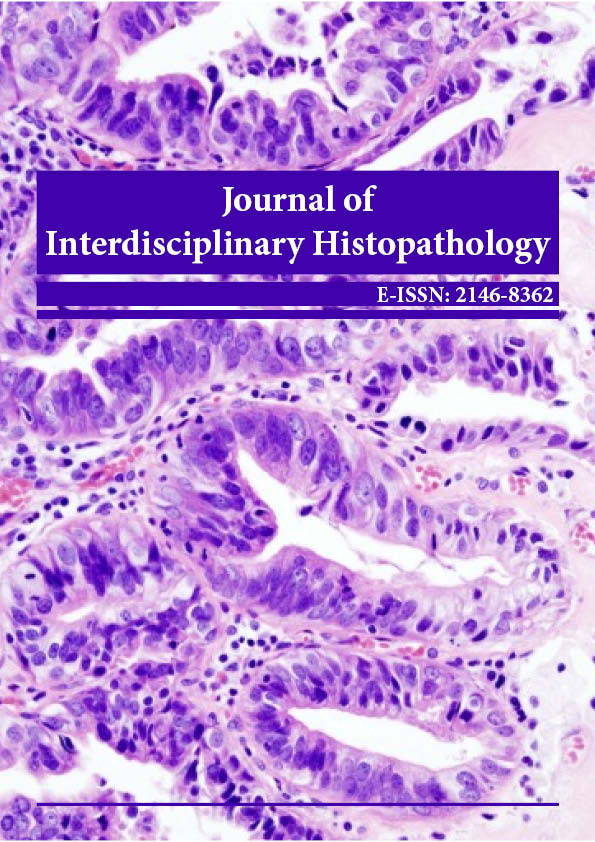
H&E-stained slides are examined under a light microscope by pathologists, histotechnologists, and researchers. The differential affinity of Hematoxylin and Eosin for various cellular components allows for the identification and evaluation of various tissue structures, such as:
Nuclei: Stained blue/purple, providing information about cell density, size, and shape.
Cytoplasm: Stained pink/red, helping to assess the amount and distribution of cellular cytoplasm.
Extracellular matrix: Stained pink/red, indicating the presence of connective tissues and their variations.
Inflammation: Characterized by the presence of immune cells, granulomas, or inflammatory infiltrates.
Limitations and advancements: While Hematoxylin and Eosin staining is a powerful and versatile technique, it does have limitations. It is not specific to certain cell types or pathological conditions, and additional staining techniques, such as immunohistochemistry, are often required for a more precise diagnosis.
In recent years, advances in staining methods and imaging technologies have expanded the capabilities of histopathology. Multiplex staining, digital imaging, and artificial intelligence-assisted analysis are some of the innovations enhancing the accuracy and efficiency of histological examinations.
Hematoxylin and Eosin staining remains the cornerstone of histopathology, providing invaluable insights into tissue architecture and cellular morphology. Its widespread use and compatibility with routine laboratory equipment have made it an essential technique for pathologists worldwide. As technology continues to evolve, Histopathologists eagerly await further advancements that will undoubtedly refine and augment the already remarkable capabilities of this vital staining method.
Copyright: © 2023 The Authors. This is an open access article under the terms of the Creative Commons Attribution Non Commercial Share Alike 4.0 (https://creativecommons.org/licenses/by-nc-sa/4.0/). This is an open access article distributed under the terms of the Creative Commons Attribution License, which permits unrestricted use, distribution, and reproduction in any medium, provided the original work is properly cited.


