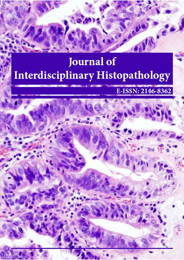
Commentary - Journal of Interdisciplinary Histopathology (2023)
Management of Hepatocytic Ballooning, Mechanisms and its Diagnosis
Joshua Ranstam*Joshua Ranstam, Department of Pathology, University of Gothenburg, Gothenburg, Sweden, Email: ranstam@gmai.com
Received: 24-Nov-2023, Manuscript No. EJMJIH-23-122919 ; Editor assigned: 27-Nov-2023, Pre QC No. EJMJIH-23-122919 (PQ); Reviewed: 12-Dec-2023, QC No. EJMJIH-23-122919 ; Revised: 20-Dec-2023, Manuscript No. EJMJIH-23-122919 (R); Published: 28-Dec-2023
Description
Hepatocytic ballooning is a term used in the field of pathology to describe a morphological change observed in liver cells, specifically hepatocytes. This alteration is often associated with various liver diseases, particularly Non-Alcoholic Fatty Liver Disease (NAFLD) and its more severe form, Non- Alcoholic Steato Hepatitis (NASH). Understanding hepatocytic ballooning is essential for clinicians and researchers, as it serves as a histological marker of liver injury and is closely linked to the progression of liver diseases [1-4].
To comprehend hepatocytic ballooning, it’s crucial to first understand the basic structure and function of hepatocytes. Hepatocytes are the primary functional cells of the liver and play a vital role in processes such as metabolism, detoxification, and the synthesis of proteins. In the context of liver diseases, hepatocytic ballooning refers to a distinct change in the shape and size of these hepatocytes [5].
When hepatocytes undergo ballooning, they become enlarged and round in shape, often resembling balloons. This morphological alteration is indicative of cellular stress and injury. Hepatocytic ballooning is typically accompanied by other histological features, including the presence of cytoplasmic vacuoles, which are often filled with lipids, and alterations in the nucleus [6].
The association between hepatocytic ballooning and non-alcoholic fatty liver disease is particularly notable. In the early stages of NAFLD, there is an accumulation of fat within hepatocytes, a condition known as hepatic steatosis. While hepatic steatosis itself may not cause significant liver damage, the progression to NASH involves additional factors, and hepatocytic ballooning becomes a prominent feature [7-9].
The exact mechanisms leading to hepatocytic ballooning are complex and multifactorial. Oxidative stress, mitochondrial dysfunction, and inflammation are among the factors implicated in the development of ballooning degeneration. As hepatocytes become stressed due to various insults, their normal cellular functions are compromised, leading to the morphological changes associated with ballooning [10].
In the context of NAFLD and NASH, hepatocytic ballooning is considered a key histological feature associated with disease severity. NASH is characterized by hepatic inflammation and hepatocellular injury, and the presence of ballooning degeneration is often a critical factor in determining the stage and prognosis of the disease. Hepatocytic ballooning is commonly assessed alongside other histological features, such as lobular inflammation and fibrosis, in liver biopsies to diagnose and stage the severity of NAFLD and NASH.
The clinical significance of hepatocytic ballooning extends beyond NAFLD and NASH. It is also observed in other liver diseases, including viral hepatitis, alcoholic liver disease, and drug-induced liver injury. In each of these conditions, hepatocytic ballooning is indicative of ongoing liver damage and can aid in the diagnosis and management of these diseases.
Researchers and clinicians employ various diagnostic techniques to detect and evaluate hepatocytic ballooning. Liver biopsy remains the gold standard for assessing histological features, including ballooning degeneration. Non-invasive imaging techniques, such as electrography and magnetic resonance imaging (MRI), are also used to assess liver stiffness and fat content, providing additional information about liver health and potential injury.
The clinical management of hepatocytic ballooning often involves addressing the underlying causes
of liver injury. For example, in the context of NAFLD and NASH, lifestyle modifications such as weight loss, dietary changes, and increased physical activity are commonly recommended. In more advanced cases, pharmacological interventions may be considered to manage specific aspects of the disease, such as insulin resistance or inflammation.
In conclusion, hepatocytic ballooning is a histological feature that signifies cellular stress and injury in the liver. While it is closely associated with non-alcoholic fatty liver disease, particularly its progressive form, non-alcoholic SteatoHepatitis, hepatocytic ballooning is also observed in various other liver diseases. Understanding and recognizing hepatocytic ballooning are crucial for clinicians and pathologists as it aids in the diagnosis, staging, and management of liver diseases. Ongoing research in this field continues to enhance our understanding of the underlying mechanisms and potential therapeutic strategies to address hepatocytic ballooning and its associated liver pathologies.
References
- Rahib L, Wehner MR, Matrisian LM, Nead KT. Estimated projection of US cancer incidence and death to 2040. JAMA Network Open. 2021;4(4):e214708.
[Crossref][Google Scholar][PubMed].
- Bengtsson A, Andersson R, Ansari D. The actual 5-year survivors of pancreatic ductal adenocarcinoma based on real-world data. Sci Rep. 2020;10(1):16425.
[Crossref][Google Scholar][PubMed].
- Morani AC, Hanafy AK, Ramani NS, Katabathina VS, Yedururi S, Dasyam AK, et al. Hereditary and sporadic pancreatic ductal adenocarcinoma: Current update on genetics and imaging. Radiol Imaging Cancer. 2020;2(2):e190020.
[Crossref][Google Scholar][PubMed].
- Aslanian HR, Lee JH, Canto MI. AGA clinical practice update on pancreas cancer screening in high-risk individuals: expert review. Gastroenterology. 2020;159(1):358-362.
[Crossref][Google Scholar][PubMed].
- Al-Shaheri FN, Alhamdani MS, Bauer AS, Giese N, Buchler MW, Hackert T, et al. Blood biomarkers for differential diagnosis and early detection of pancreatic cancer. Cancer Treat Rev. 2021;96:102193.
[Crossref][Google Scholar][PubMed].
- Petrov MS, Yadav D. Global epidemiology and holistic prevention of pancreatitis. Nat Rev Gastroenterol Hepatol. 2019;16(3):175-184.
[Crossref][Google Scholar][PubMed].
- Kirkegard J, Cronin-Fenton D, Heide-Jørgensen U, Mortensen FV. Acute pancreatitis and pancreatic cancer risk: a nationwide matched-cohort study in Denmark. Gastroenterology. 2018;154(6):1729-1736.
[Crossref][Google Scholar][PubMed].
- Ouyang G, Pan G, Liu Q, Wu Y, Liu Z, Lu W, et al. The global, regional, and national burden of pancreatitis in 195 countries and territories, 1990-2017: A systematic analysis for the Global Burden of Disease Study 2017. BMC Med. 2020;18:1-3.
[Crossref][Google Scholar][PubMed].
- Keene BW, Atkins CE, Bonagura JD, Fox PR, Haggstrom J, Fuentes VL, et al. ACVIM consensus guidelines for the diagnosis and treatment of myxomatous mitral valve disease in dogs. J Vet Intern Med. 2019;33(3):1127-1140.
[Crossref][Google Scholar][PubMed].
- Gaasch WH, Meyer TE. Left ventricular response to mitral regurgitation: implications for management. Circulation. 2008;118(22):2298-2303.
[Crossref][Google Scholar][PubMed].
Copyright: © 2023 The Authors. This is an open access article under the terms of the Creative Commons Attribution Non Commercial Share Alike 4.0 (https://creativecommons.org/licenses/by-nc-sa/4.0/). This is an open access article distributed under the terms of the Creative Commons Attribution License, which permits unrestricted use, distribution, and reproduction in any medium, provided the original work is properly cited.


